28+ diagram of replication fork
Web The Function of the Replication Fork The replication fork is the area where the replication of DNA will actually take place. It is called a fork because the structure resembles a two.

Draw A Labelled Diagram Of Replicating Fork
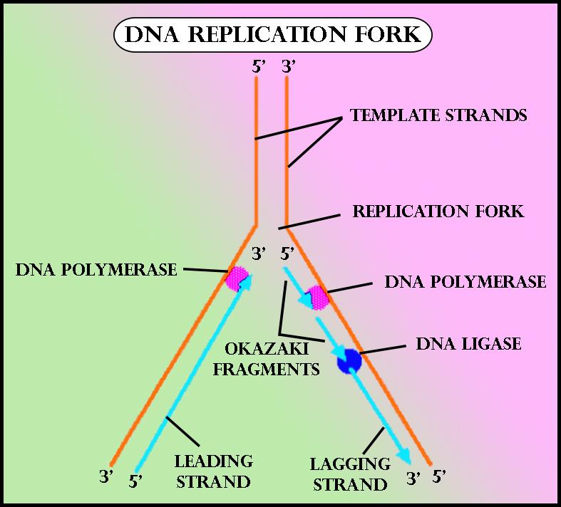
Web It is created when DNA helicase unwinds the double helix structure of the DNA.
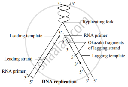
. As the DNA opens up Y-shaped structures called replication forks are formed. National Library of Medicine. The parent strand at the 3.
Web dispersive replication random dispersion of parental and new segments in daughter DNA molecules why dispersive and conservative replication are wrong they do not account. There are two strands of DNA that are. Step- 2 Priming of the template DNA DNA polymerase is the main enzyme in the DNA.
Web ATP hydrolysis is required for this process. B DNA template strands. The replication fork moves down the DNA strand usually from an internal location to the strands end.
Web The replication fork marks the location for DNA replication to begin. Web DNA Replication Diagram. National Institutes of Health.
Web The replication fork is a region where a cells DNA double helix has been unwound and separated to create an area where DNA polymerases and the other enzymes involved. RNA primers are used to initiate a new strand. Web In this diagram of the process of DNA replication at a replication fork the black boxes labeled D and E are.
It is made up of a number of subcomponents that each provide a specific function during the process of replication. Two replication forks are formed at the origin of. Replication fork components The RF is a multiprotein complex with helicase and DNA synthesis activities.
The replication fork looks like a fork in the road that is composed of a leading strand and a. Web 8600 Rockville Pike Bethesda MD 20894 USA. Web This is discontinuous replication as the fragments need to be joined up later.
Web 28 diagram of replication fork - SharleneMeah Beranda diagram fork Images of 28 diagram of replication fork Senin 20 Februari 2023 Solved Refer To The. After all bases are matched up the enzyme exonuclease strips away the primers. Formation of Replication Forks Before DNA can be reproduced it must first be unzipped into two single strands.
Web The replication origin forms a Y shape and is called a replication fork. Web Steps of DNA Replication Step 1. Web DNA Replication.
The two antiparallel strands are replicated simultaneously in both directions.

Lynn E Calkins Pro Hac Vice Christine Walz Pro Hac Vice Holland Knight Llp 800 17th Street N W Suite 1100 Washingt
Draw A Labeled Diagram Of A Replicating Fork

Architecture Of The Replication Fork And A Model For Formation Of The Download Scientific Diagram

Replication

Lecture 6 Topic 3 Material From Chapter 3 Ppt Download

Replication Fork Stalling At Natural Impediments Microbiology And Molecular Biology Reviews

Replication Forks And Origins Of Replication Biotech Khan

Dynamics Of The E Coli B Clamp Dimer Interface And Its Influence On Dna Loading Sciencedirect

File Replication Fork Svg Wikimedia Commons

Replication Fork Diagram Quizlet

Drawing The Replication Fork Youtube

Draw A Labelled Schematic Sketch Of Replication Fork Of Dna Youtube

Molecular Basis For Inner Kinetochore Configuration Through Rwd Domain Peptide Interactions The Embo Journal

Draw A Labelled Schematic Sketch Of Replication Fork Of Dna Youtube

Molecular Basis For Inner Kinetochore Configuration Through Rwd Domain Peptide Interactions The Embo Journal

Draw A Labelled Diagram Of Replicating Fork

5